Confirm before you fuse.

Posterior SI joint fusion

Hold your permanent SI joint implants to the exact same standard as your diagnostic injections.

Patented. U.S. Patent No. 12,472,070. Patent pending, US 2026/0069421 A1. Other patents pending.

Looking for patient information?

The SI joint fools skilled hands.

22%

of landmark guided SI injections actually reached the joint in a CT verified series.1

49%

of needles that felt intra articular under fluoroscopy were confirmed in the joint by contrast.2

98%

with fluoroscopy and contrast. Contrast found the rest, and the needle was moved.3

If you verify the injection, why wouldn’t you verify the implant? A diagnostic SI joint injection means nothing unless contrast proves it’s in the joint. UNIO CONFIRM™ brings that same standard of precision and accuracy to permanent fusion.

Request the technique guide

1. Rosenberg JM, Quint TJ, de Rosayro AM. Clin J Pain. 2000;16(1):18. PMID 10741814. Landmark guided SI injections localized by CT: 8 of 37 (22%) intra articular; epidural injectate in 24%.

2. Nacey NC, Patrie JT, Fox MG. AJR Am J Roentgenol. 2016;207(5):1055. Fluoroscopically guided SI joint injections; intra articular placement 49% when classified only by contrast clearly within the joint.

3. Jee H, et al. Arch Phys Med Rehabil. 2014;95(2):330. Randomized, 110 patients; fluoroscopy 98.2% vs ultrasound 87.3% intra articular by contrast; fluoroscopy first pass 92%.

Injection studies describe diagnostic needle placement, not fusion implants. No head to head study with UNIO® is implied.

Why confirm before you fuse

Fluoroscopy alone can miss the joint.

SI joint anatomy varies in roughly one of every three to four patients, and in far more in some series.4

A 2025 posterior SI joint fusion series concluded that intraoperative fluoroscopy may not be sufficient to find the target, and called preoperative CT or MRI mapping essential.4

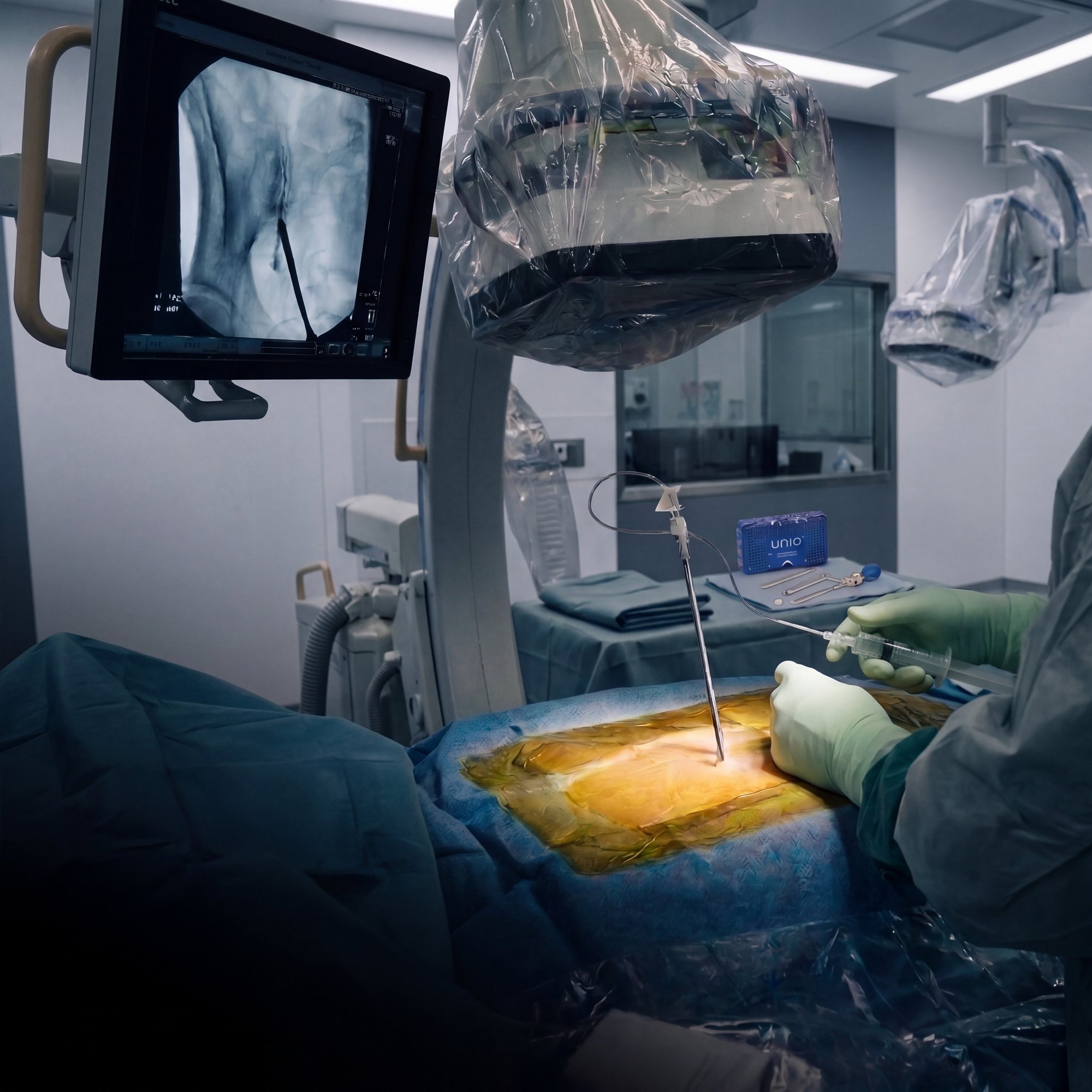
Mapping plans the path. Contrast proves it. UNIO CONFIRM™ shows intra articular placement with live contrast through the pin, on the table, before the joint is prepared and before anything permanent is implanted.

What happens when the implant misses
37

posterior SI joint fusion cases that failed and went to revision surgery, across seven centers.5

0

showed radiographic fusion of the SI joint before revision.5

Outside the joint

Suboptimal implant position was a recurring finding. The authors reported many implants placed outside the joint.5,6

The question the authors left open

“How often does extraarticular placement occur?”6

The UNIO® answer

Know before you implant. UNIO CONFIRM™ shows the path is intra articular with live contrast through the pin, in every case, before the joint is prepared.

The problem is written into our patent
“Many procedures are complicated, and use complex instrumentation, and when using a posterior approach to enter the SI joint, sometimes result in the implant being placed in an improper location.”
U.S. Patent No. 12,472,070, specification.

4. Bovinet C, Moghim R, Jin MY, Abd-Elsayed A. Interv Pain Med. 2025;4(2):100581. PMID 40242295. Single center retrospective series, 208 patients, posterior allograft SI joint fusion with preoperative mapping. Reported prevalence of SI joint anatomical variation in most cited studies: 25.7% to 35.7%.

5. Kranenburg A, et al. Med Devices (Auckl). 2022;15:229. PMID 35899066. Retrospective case series of 37 patients revised after failed posterior standalone structural allograft. A series of failures cannot establish how often malposition occurs. Authors include employees of a lateral implant manufacturer.

6. Kranenburg A, et al. Author reply. Med Devices (Auckl). 2022;15:367. PMID 36281345.

These studies did not evaluate UNIO®. No head to head study is implied.

Two audiences. One standard of precision.

For interventional pain management

You would not trust a diagnostic SI joint injection without contrast confirmation; the result means nothing if the anesthetic misses the target. UNIO CONFIRM™ lets you bring that exact same diagnostic certainty to your permanent fusion procedures.

For spine surgeons

Prove intra articular position before you prepare the joint, then decorticate both surfaces for a true biological arthrodesis. UNIO CONFIRM™ combines rigorous validation with real bone and the fixation to back it when the joint needs it.

The UNIO CONFIRM™ method

Confirm before you fuse.

01

Confirm.

A blunt, cannulated pin safely accesses the inferior joint. Live image contrast injection through its lumen provides definitive, intra articular placement.

02

Decorticate.

The Access Portal seats along the verified path. A hand drill through its two channels decorticates both the sacral and iliac surfaces. True joint preparation is what drives the fusion process.

Why it matters Moves past simple stabilization to achieve true joint arthrodesis.

03

Unite.

The UNIO® SI Graft, a structural cortical allograft preloaded with UNIO DBM™, is advanced into the prepared joint. Its sharp, directional teeth engage bone and pierce the lateral cortex of the sacrum and the medial cortex of the ilium, seating the implant across the joint. Real bone and the patient’s own biology finish the fusion.

Why it matters Integral, self retaining fixation and natural bone remodeling in one implant, with titanium fixation available when the joint needs it.

A physician in a mask and protective eyewear, focused during a procedure
Intellectual property

Issued and pending.

Patents and applications owned by Resilient Surgical Holdings, Inc. Claim language reproduced in full. Full notice on the patents page.

A joint stabilization method comprising: placing an elongated blunt-tipped pin within a joint of a human, the blunt-tipped pin including a cannulated interior; placing at least one of contrast and medicine within the cannulated interior of the elongated blunt-tipped pin; placing a dilator having a substantially hollow interior over the elongated blunt-tipped pin, while the elongated blunt-tipped pin remains inserted within the joint; placing a drill guide over the dilator and the elongated blunt-tipped pin, the drill guide having a substantially hollow interior section; removing the dilator and the elongated blunt-tipped pin, leaving the drill guide; drilling through the interior of the drill guide to provide a pathway for an implant; and placing the implant within the pathway, the implant to reside within the joint.

Patent pending. U.S. Patent Application Publication No. US 2026/0069421 A1, Application No. 19/391,864, claim 11

A method comprising: placing an elongated pin within a joint of a biologic organism; placing a dilator having a substantially hollow interior over the elongated pin, while the elongated blunt-tipped pin remains inserted within the joint; placing a drill guide over the dilator and the elongated blunt-tipped pin, the drill guide having a substantially hollow interior section; removing the dilator and the elongated pin, leaving the drill guide; drilling through the interior of the drill guide to provide a pathway for an implant; and placing the implant within the pathway, the implant to reside within the joint.

Patent pending. U.S. Patent Application Publication No. US 2026/0069421 A1, Application No. 19/391,864, claim 8

A blunt-tipped pin used for stabilization of an SI Joint, the elongated blunt-tipped pin comprising: an elongated body having a substantially cannulated interior portion, the elongated body having a first open end, the first open end configured to receive at least one of contrast and medicine, the elongated shaft having a second end configured to probe the SI joint.

Patent pending. U.S. Patent Application Publication No. US 2026/0069421 A1, Application No. 19/391,864, claim 1

A system for stabilizing a human joint, the system comprising: an elongated blunt-tipped pin configured to be inserted within a joint of a human, the blunt-tipped pin including a cannulated interior; a dilator having a substantially hollow interior configured to be placed over the elongated blunt-tipped pin, while the elongated blunt-tipped pin remains inserted within the joint; and a drill guide having a substantially hollow interior configured to be placed over the dilator and the elongated blunt-tipped pin, the drill guide configured to receive at least a portion of a drill for providing a pathway for an implant.

One method. Every site of service.

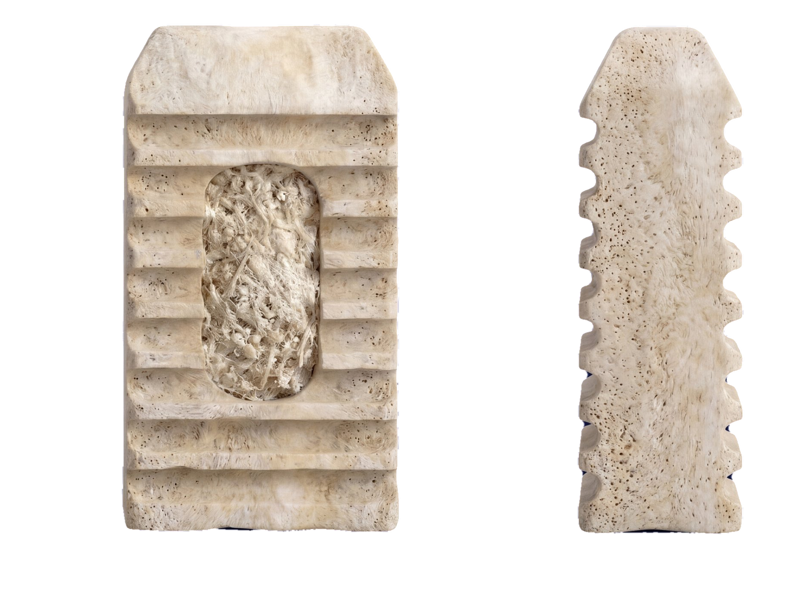
UNIO SI implant shown from the front and from the side with dimensions: 22.5 mm tall, 12 mm wide, 7 mm thick
UNIO® SI Graft
Surgery center, hospital and OBL

Structural cortical allograft, 22.50 mm by 12 mm by 7 mm, placed through a reusable tray. The flagship. A central graft window carries UNIO DBM™. Both faces carry integral, self retaining mechanical fixation elements: a series of sharp, directional, barbed teeth machined into the bone, which engage bone and pierce the lateral cortex of the sacrum and the medial cortex of the ilium as the implant seats across the joint. The lateral faces are smooth. The tapered end leads; the flat end faces posterior.

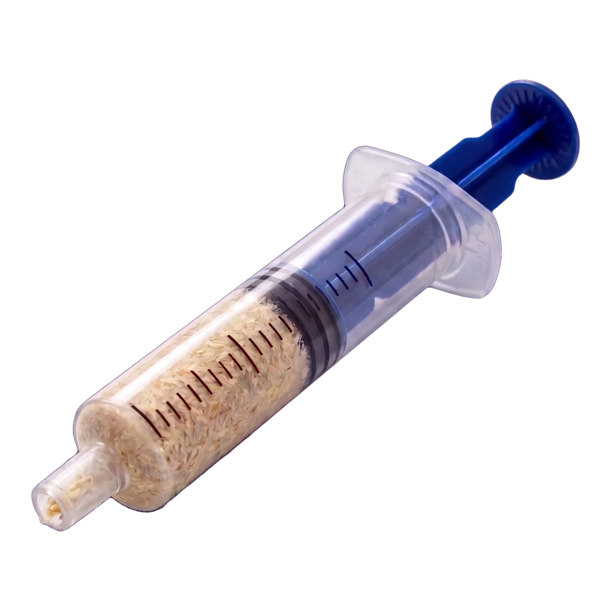
A syringe of UNIO DBM demineralized bone matrix on a sterile field
UNIO DBM™
Biologic

Demineralized bone matrix, packed into the implant window before placement. Allograft, like the implant it fills.

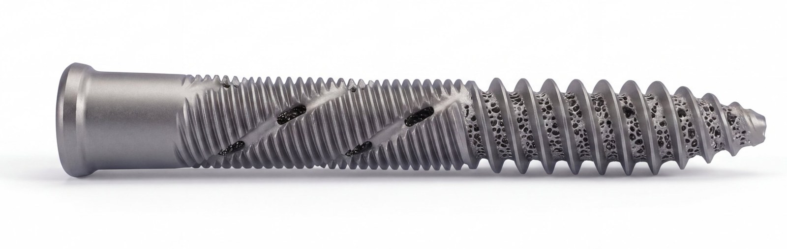
UNIO SI Ti™
When the joint needs it

Titanium fixation for permanent mechanical stabilization. Biologic when possible. Metal when necessary.

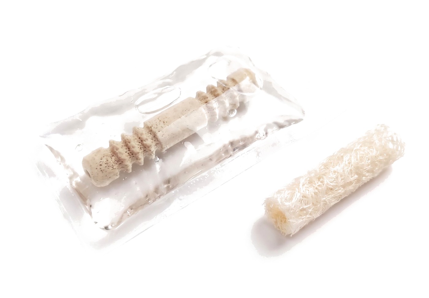
UNIO BioFuze in its sterile pouch with the demineralized bone fiber jacket beside it
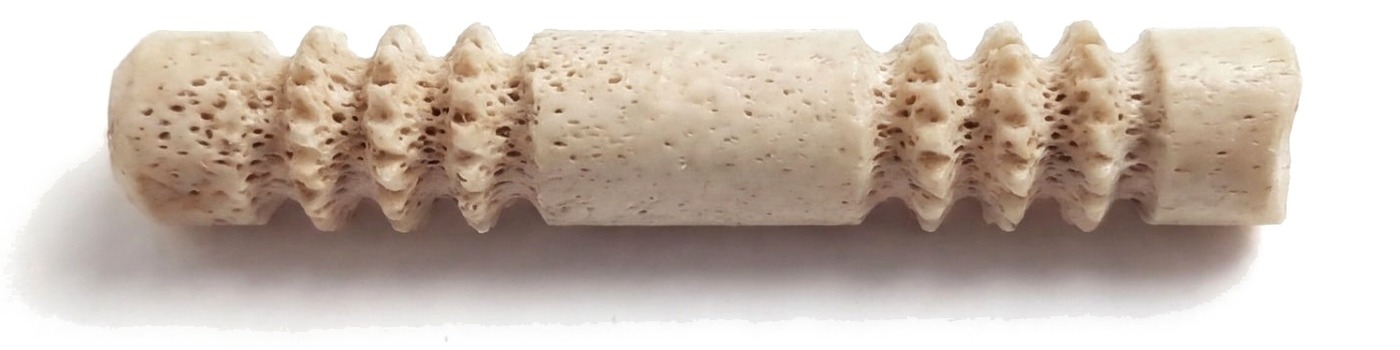
UNIO BioFuze Graft, the cortical allograft with intrinsic fixation
UNIO BioFuze™ Graft
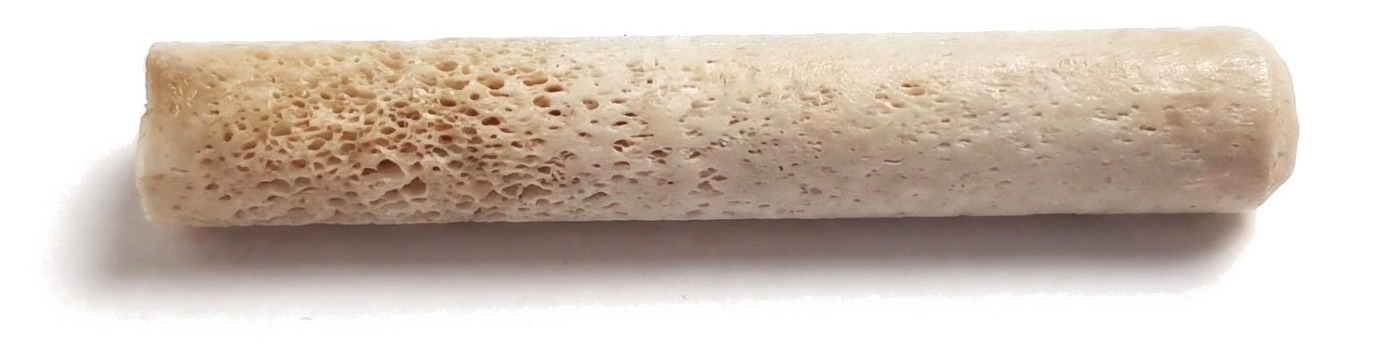
UNIO BioFuze Dowel, the smooth cortical allograft
UNIO BioFuze™ Dowel
UNIO BioFuze™

Fully disposable, percutaneous.

Schedule a demo
Technique guide and ordering →Instructions for Use →
Every site of service

Reusable where it saves. Disposable where it counts.

The technique does not change with the room. The format does. Physicians trained once carry the same method from the office to the surgery center, and escalating a patient never means changing systems.

Surgery center and hospital

Consigned reusable tray, no capital outlay. Predictable per case cost at any volume. One vendor, one method, one training track.

Office and procedure suite

Fully disposable configuration. No sterile processing, no tray turn, no case delay waiting on reprocessing.

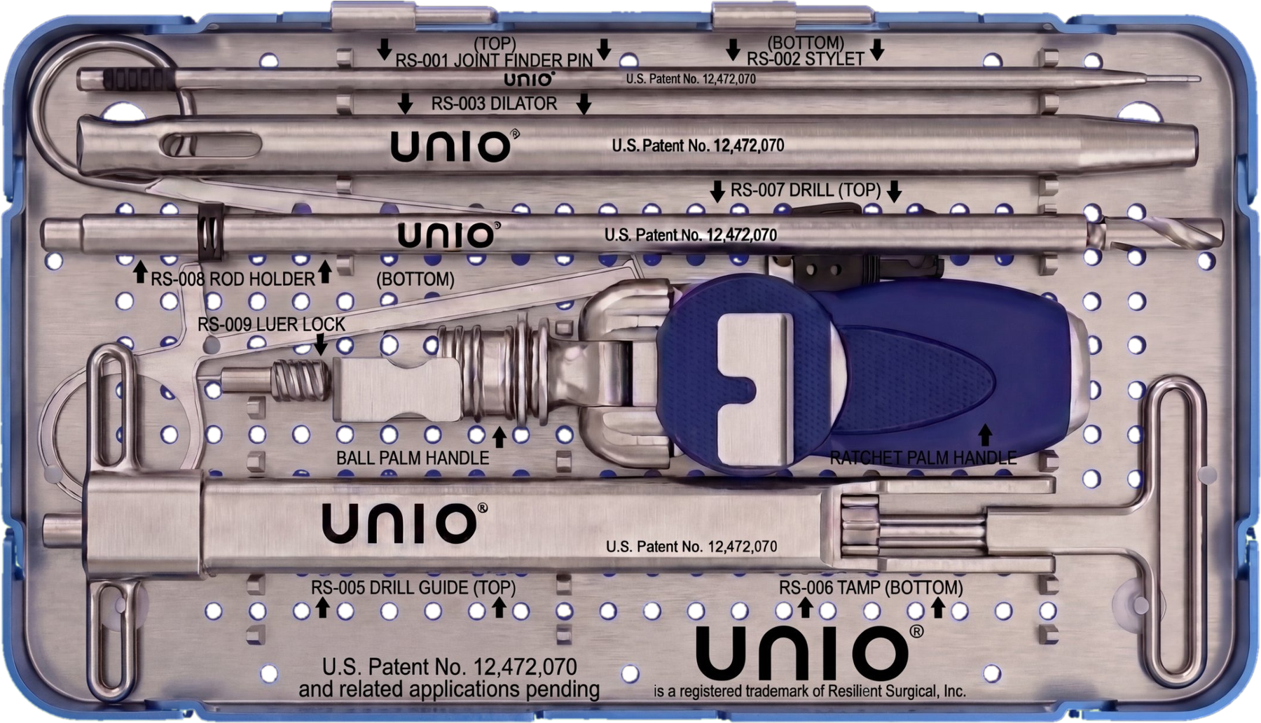
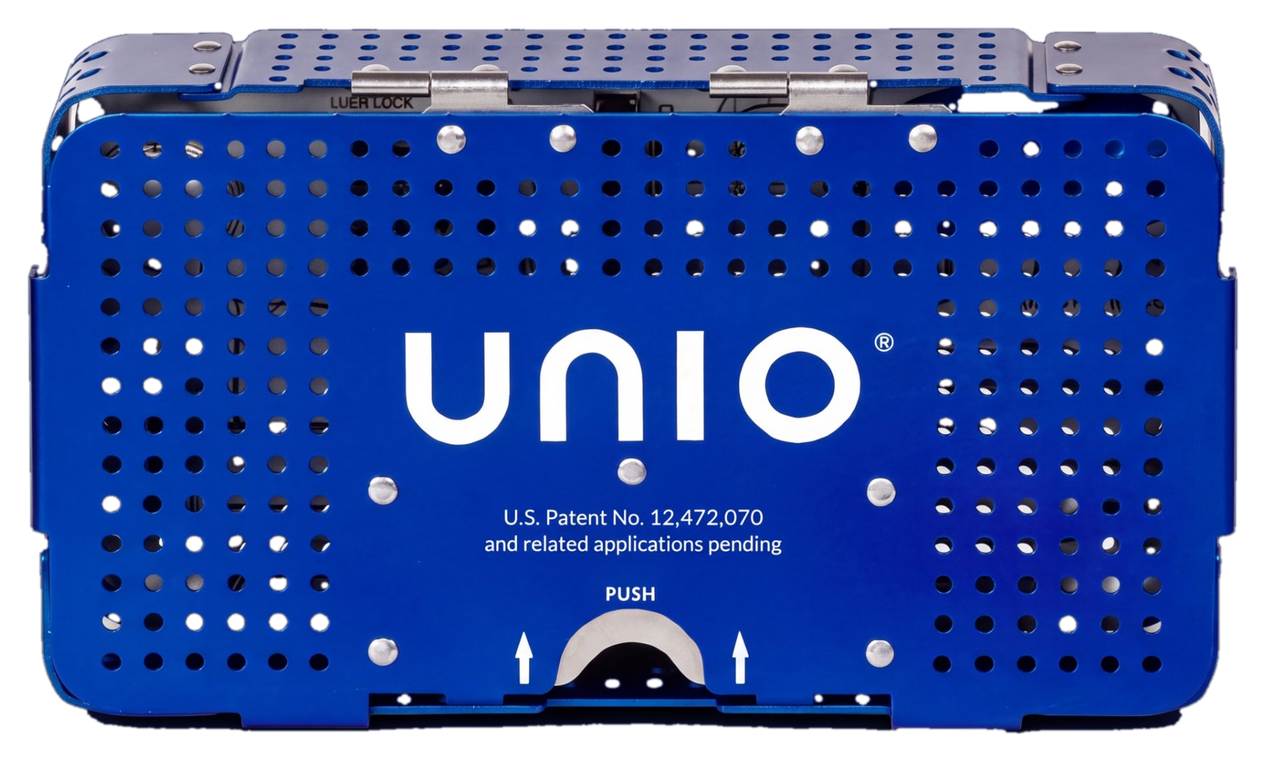
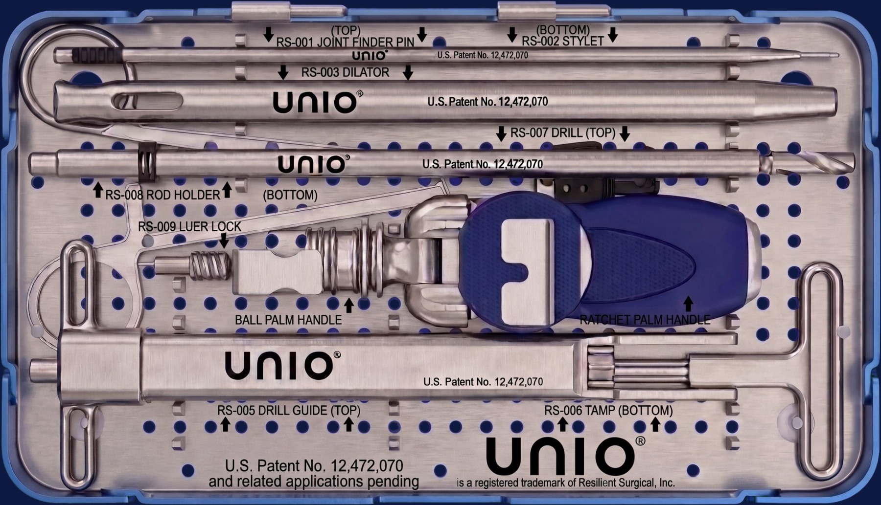
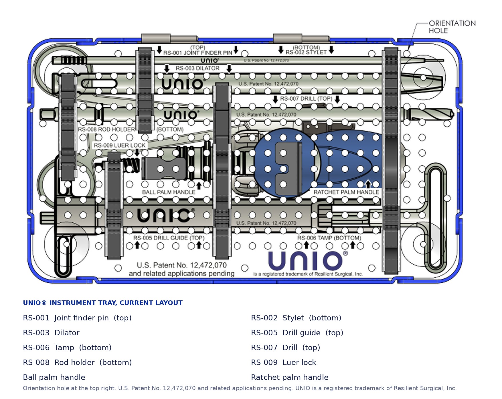
The open UNIO tray with the instrument set in its silicone brackets The closed UNIO case with the UNIO logo and the patent marking on the lid Hover to open the trayTap to open the tray
The reusable set

One tray carries the method in the surgery center, the hospital and the OBL. Marked with U.S. Patent No. 12,472,070.

Where you will find us

Societies and meetings we support.

Sponsor, exhibitor, or faculty at the medical, pain, and surgical meetings where posterior SI fusion is taught, advanced, and debated.

In Georgia, the SBWC Annual Educational Conference brings together the state’s adjusters, case managers, and medical directors. Contrast confirmed placement gives them what utilization review asks for: objective documentation that the joint treated is the joint that was diagnosed.

Request a cadaver lab and peer discussion

Evaluate the UNIO CONFIRM™ method firsthand.

See the difference that contrast verified precision and biological fusion make. Choose how you want to experience the system.

01

Request a bioskills lab

Secure a spot at an upcoming local or regional cadaver lab to experience the cannulated delivery system and tactile decortication workflow firsthand.

Request lab access
02

Speak with a physician

Connect directly with an active spine surgeon or interventional pain specialist currently utilizing the UNIO® system in their practice.

Speak with a physician
Bone first. Metal when it is needed.

The UNIO® system gives you the flexibility to adapt directly to what the joint dictates. Both pathways deploy over the exact same confirmed pin.

Surgeon FAQs and learning curve

Straight answers from training and the first cases. Ask us anything these do not cover.

What is the learning curve for transitioning from a lateral metal screw system to the UNIO® posterior/oblique approach?

The learning curve is minimal because the system relies on familiar anatomical landmarks and fluoroscopic views. For IPM physicians, the initial step mimics a standard diagnostic SI joint injection.

How much time does the contrast confirmation step add to the standard OR timeline?

It adds virtually no time. Because the pin is cannulated, connecting a syringe and confirming contrast flow under live imaging takes less than 60 seconds in our training experience. This minor step prevents the operative and clinical delays associated with malpositioned hardware.

Is the structural allograft strong enough to stabilize the SI joint immediately?

The structural cortical allograft placed across the prepared joint provides immediate mechanical stability across the joint space. When structural titanium fixation is clinically dictated by the patient’s anatomy or bone quality, the UNIO® instrumentation allows for rigid titanium screw placement over that exact same confirmed pin pathway.

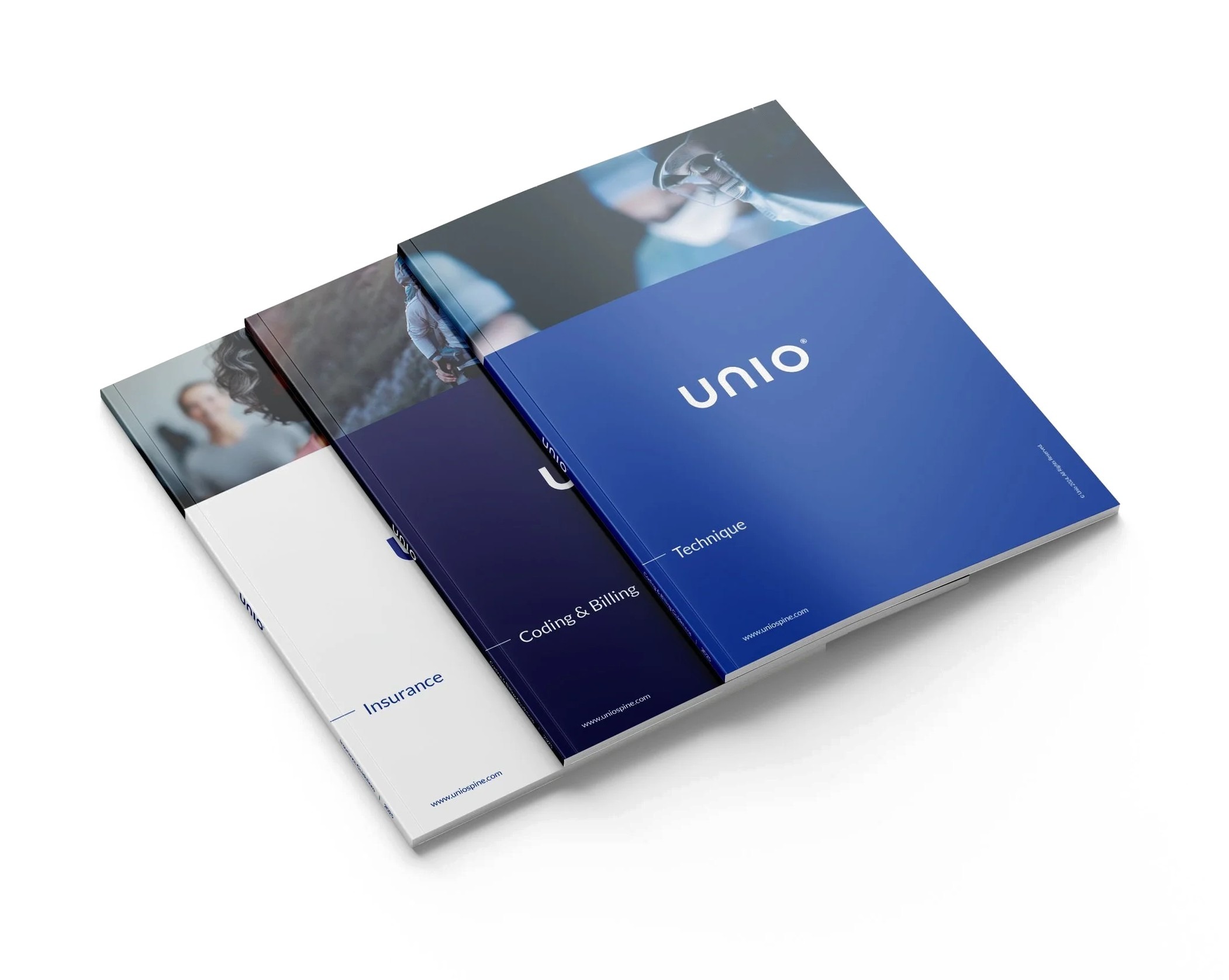
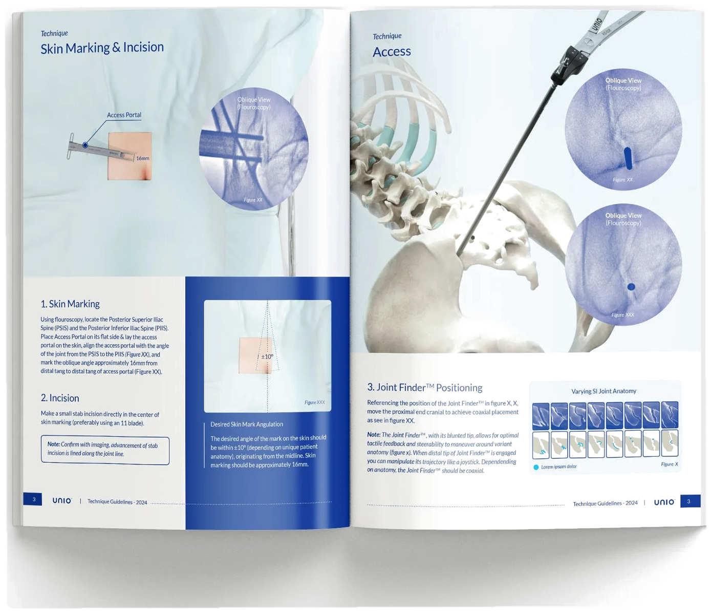
Printed guides

The blueprint your office works from.

Printed, bound and current: the technique step by step, code selection from the op note, and the payer checklists, re verified every quarter.

  • Technique. The CONFIRM method step by step and the instrument set.
  • Coding and billing. Code selection from the op note, the documentation that supports it, and the economics by site of service.
  • Insurance. Every policy we work with, what each requires, and the checklist for each payer.
The three UNIO guides, Technique, Coding and Billing, and Insurance, fanned on a white surface The technique guide open to the skin marking, incision and access pages

Bring confirmation to your practice.

Tell us what you need and the right person answers.
customerservice@uniospine.com 229 496 4493
UNIO Execute™
Documentation support and case coordination for participating UNIO® practices.